
Total Articles: 43
Today's Visitors: 40

Journal of the Bahrain Medical Society
Year 2009, Volume 21, Issue 1, Pages 197-200
Dr. Noora Al- Kobaisi1
1Department of Ophthalmology, Salmanya Medical Complex, Kingdom of Bahrain

AIM: To report the results of Bahrain diabetic retinopathy photo screening program during 2007
Methods: We audited the results of diabetic retinopathy photo screening program using digital retinal camera in Bahrain from the period January 2007 till December 2007
Results: A total of 3265 patients were screened by digital retinal camera 2359 patients (72 %) showed no diabetic retinal changes. Mild nonproliferative changes were noticed in 711 patients (22%). Moderate nonproliferative changes were noticed in 83 patients (3%) Severe nonproliferative changes were noticed in 11 patients (0.3%). Proliferative retinopathy changes were noticed in 44 patients (1 %). Maculopathy was found in 123 patients (4%).
Conclusion: Digital retinal photo screening is practical in Health Centers and it can detect the normal retina from the retinopathy changes in diabetic patients accurately. Its implementation has been associated with a reduction in presentations with vision-threatening retinopathy within the total community. The normal eyes on initial photo screening yearly repeat photo screening schedule for this group.
Keywords: Diabetic retinopathy, diabetic retinopathy screening program, diabetic screening by digital camera, Proliferative retinopathy, nonproliferative retinopathy.
Introduction
Diabetic retinopathy is a highly specific micro vascular complication of both insulin dependant (type1) and non insulin dependant (type 2) diabetes. The prevalence of retinopathy is strongly linked to the duration of diabetes. After 20 years of diabetes nearly all patients with type 1diabetes and over 60% of patients with type 2 diabetes have some degree of retinopathy. Up to a fifth of newly diagnosed diabetics have been found to have some retinopathy. A diabetic is 25 times more likely to go blind than a person in the general population. Diabetic retinopathy poses a serious threat to vision.
Study Methods
Surveillance and treatment of diabetes-related complications should be part of routine care of all patients with diabetes. Since even advanced disease can be asymptomatic. The preferred method for screening is digital retinal photography. This technology has secondary advantages of easy storage and retrieval of images, which facilitates quality assurance, training, and patient education. Several studies have reported the cost effectiveness of screening for retinopathy. They have established that screening for diabetic retinopathy saves vision at a relatively low cost.
The Screening Process
The prevalence of diabetics in Bahrain is said to be 25-30 per cent among the nationals. The aim of diabetic retinopathy screening program in BAHRAIN is to develop an integrated screening program to prevent blindness caused from diabetic retinopathy. Main focus is on early detection and management of diabetic retinopathy.
In Bahrain there are 21 Health Centers at present and this program covers only 15 Health Centers. The fully digitalized screening clinic for diabetic retinopathy for diabetic patients is established and functioning at present and covers 15 Health Centers. Muharraq health center which covers 4 health centers and A’ali which covers 11 health centers. All the patients attending the health centers with diabetes are registered and the patients are directed to the two screening centers with an appointment system.
Screening Methods
Non mydriatic digital retinal camera Canon CR-6-45 NM single field fundus photography is used to take the digital fundus pictures of the diabetic patients. Patient’s eyes were dilated with Mydryacyl 1 % drops. The digital photography is performed by ophthalmic technician trained in taking the digital fundus picture. The digital fundus pictures are electronically transmitted ophthalmology department in Salmaniya Medical Center. Consultant retina specialist routinely view the digital fundus pictures grade these images and plan the follow up or call them for direct visualization by ophthalmoscope examination and plan the modality of treatment.
Results
From January 2007 to December 2007, 3265 patients were screened for diabetic retinopathy. We found that 2359 (72%) didn’t show any retinopathy changes therefore they are rescheduled for repeat photo screening in a year.
There are two stages of diabetic retinopathy – nonproliferative and proliferative retinopathy:
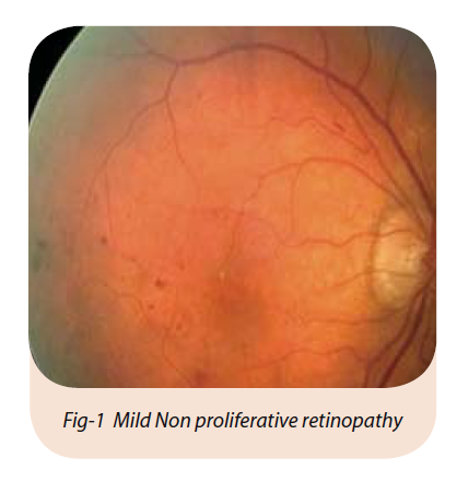
Nonproliferative retinopathy is the earlier stage. In this stage there may be hemorrhages (bleeding) in the retina with leakage of blood causing a “wet retina” or protein deposits (exudates) in the retina. As a consequence, the retina does not receive enough oxygen. This early stage of diabetic retinopathy usually produces no visual symptoms but, if there is fluid in the central portion of the eye (macular edema), vision is diminished.
Diabetic retinopathy has four stages
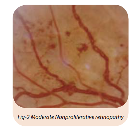
2. Moderate Nonproliferative Retinopathy. As the disease progresses, some blood vessels that nourish the retina are blocked. We found 83 patients (3 %) showed moderate nonproliferative changes in photo screening. (Fig-2)
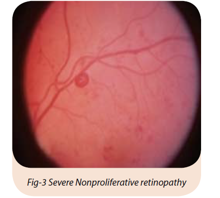
3. Severe Nonproliferative Retinopathy. Many more blood vessels are blocked, depriving several areas of the retina with their blood supply. These areas of the retina send signals to the body to grow new blood vessels for nourishment. We found 11 patients (0.3%) showed sever nonproliferative changes in photo screening.(Fig-3)
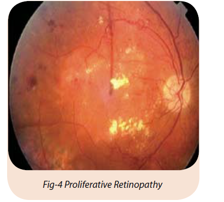
4. Proliferative Retinopathy. Proliferative retinopathy is the second stage. And more advanced stage of diabetic retinopathy,the abnormal cells rapidly spread - proliferative - across the inner surface of the retina. These weakened vessels can bleed into the vitreous. New abnormal vessels develop in the retina and grow towards the center of the eye. These vessels frequently bleed into the vitreous (the clear jelly in the center of the eye). Such bleeding episodes cause severe visual problems. Small bleeds may clear upon their own but larger bleeds need surgery. In addition, the connective scar tissue, which forms as a result of the ruptured blood vessels, can shrink, pulling the retina away from its underlying structure, causing it to detach. Severe loss of sight - even blindness - may result. We found 44 patients (1%) showed proliferative retinopathy in the photo screening. (Fig-4)
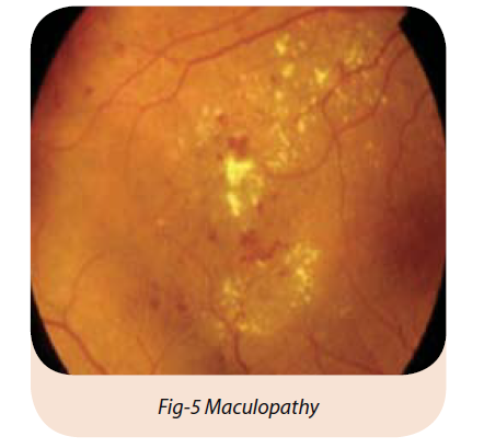
5. Maculopathy. Fluid can leak into the center of the macula, the part of the eye where sharp, ahead straight-vision occurs. The fluid makes the macula swell, blurring vision. This condition is called macular edema. It can occur at any stage of diabetic retinopathy, although it is more likely to occur as the disease progresses. About half of the people with proliferative retinopathy also have macular edema. We found 123 patients (4%) showed maculopathy in the photo screen. (Fig-5)
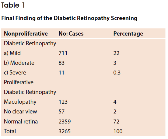
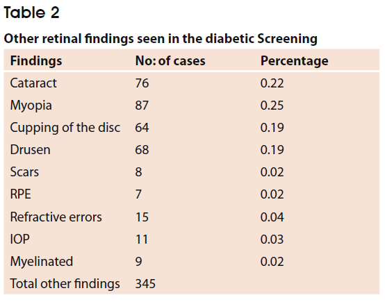
During the photo screening for diabetic retinopathy we found very significant retinal finding which are given in the following table
Discussion
It has been estimated that the total number of people with diabetes in Bahrain is 20-25 %.The explosive growth of the diabetic population demands greater efficiencies in the management of our patients with potential or actual vision-threatening condition.
A screening programme should aim to detect patients at risk when they can still be effectively treated, and this can be achieved by regularly checking the patients’ eyes. The service planning guidelines for diabetes recommended regular eye review for all patients with diabetes. This was not occurring due to the fact that comprehensive systematic review would put on the service. Our digital retinal
Photo screening program reduced the burden of normal eye screening on our ophthalmology service and allowed communitybased capture of retinal images, ensuring coverage of those subjects who do not regularly attend hospital clinics. Images were then assessed centrally by specialist ophthalmologists.
Various methods of screening have been shown to be sufficiently sensitive and specific for the detection of sight -threatening eye disease (STED) 1, 2 at justifiable costs. 3, 4 However, the sensitivity and specificity of a screening program are not the only important considerations. The extent of the population coverage and the screening intervals are vital to the success of a screening program. The digital retina screening service makes the screening more accessible to a larger number of patients.
The Diabetic Retinopathy fundus digital camera screening in Bahrain is very accurate in differentiating from normal retina to abnormal diabetic retinopathy changes. Our digital retinal photo screening program has achieved good ascertainment of the estimated diabetic population for at least one photo screen. Approximately 72% of the diabetic population in Bahrain were screened one or more times.
The percentage of the non-assessable images was 52 patients 2 %. The British Diabetic Association recommends a maximum failure rate of 5% for any screening program to be acceptable. 5, It is important to differentiate between technical reasons for poor photography and physical causes such as cataract. The photograph should be repeated if possible if the reason is technical. Overall, the number of non-assessable images has been acceptable.
We found that 2359 eyes (72.0%) didn’t show any retinal lesion and, therefore, these patients were rescheduled for repeat photo screening in one year, thus reducing the waiting list for a specialist ophthalmologist examination. The patients referred for specialist review Mild nonproliferative retinopathy 6 months, moderate at 4 months and sever at 2 monthly for direct ophthalmoscope examination with necessary treatment initiated as early as possible.
Proliferative retinopathy patients are called immediately for direct ophthalmoscopic examination and necessary treatments were initiated.
The true prevalence of sight-threatening diabetic retinopathy is unknown, current evidence suggests that the diabetic population have some diabetic retinopathy. Our data provide an estimate of the baseline prevalence of grades of diabetic retinopathy in Bahrain diabetic population, a community-based retinopathy screening program.
Significant retinopathy are followed in specialist ophthalmology clinics and do not appear on the photo screening register once they have been referred. We do not know precisely how many patients are in the specialist clinic program at the moment, but expect this to be a significant number. We intend to identify these patients to enable us to assess the percentage of expected diabetic patients in Bahrain.
In conclusion: The Diabetic Retinopathy digital retinal camera Photo screening Program has shown that digital retinal photography is a practical and effective method of screening for diabetic eye disease in Health Centers in Bahrain.
1. Harding SP, Broadbent DM, Neoh C, et al. Sensitivity and specificity of photography and direct ophthalmoscopy in screening for sight threatening eye disease: the Liverpool Diabetic Eye Study. BMJ 1995;311:1131–5.
2. Leese GP, Tesfaye S, Dengler-Harles M, et al. Screening for diabetic eye disease by optometrists using slit lamps. J R Coll Physicians Lond 1997;31:65–9.
3. James M, Turner DA, Broadbent DM, et al. Cost effectiveness analysis of screening for sight threatening diabetic eye disease. BMJ 2000;320: 1627–31.
4. Javitt JC, Aiello LP, Chiang Y, et al. Preventive eye care in people with diabetes is cost-saving to the federal government. Implications for healthcare reform. Diabetes Care 1994;17:909–17.
5. British Diabetic Association. Retinal photography screening for diabetic eye. disease: a BDA report. London: British Diabetic Association; 1997.